

Why Perform a Necropsy on Your Parrot
The objective of this article is to become familiar with this unique medical discipline, to discover the various aspects of a necropsy and its numerous benefits.
A necropsy is a generally complete study of a body after death. It is one of the oldest medical procedure, but nonetheless still unknown to most people. A necropsy is an important clinical diagnostic tool, very often this procedure will help determine the cause of death or the cause of a multifaceted disease.
The loss of our loving little feathered treasure or the unexplained loss of several birds of a breeding stock representing a lifetime’s achievement can be an emotionally painful experience. When the time comes to suggest a necropsy, the situation is even more unpleasant. It is of course difficult to accept that our precious animal will undergo that type of procedure, especially if the importance of a necropsy is not always obvious. But is it really necessary to accept a necropsy when it is suggested?
As unpleasant as this can be, a necropsy can provide us with the last crucial piece of a complex puzzle. Furthermore it can more or less provide us with an essential diagnostic tool for the future health and survival of an entire stock.
The objective of this article is to become familiar with this unique medical discipline, to discover the various aspects of a necropsy and its numerous benefits. A necropsy is an important clinical diagnostic tool, very often this procedure will help determine the cause of death or the cause of a multifaceted disease.
Potential Indications of a Necropsy (Table # 1)
Some diseases can be prevented once they are correctly identified. Among other things, we can identify infections, nutritional disorders, hereditary diseases and exposures to a toxic agent or to a physically harmful circumstance. These diseases are not always evident before death. A necropsy is the last opportunity to shed some light on them and can prevent other deaths with the help of an appropriate treatment or adequate management.
Animal infections that can be transmitted to human beings (zoonosis) can be very dangerous. Such a discovery will have important implications for the people exposed to the infected animal. It will be suggested to them to consult a doctor. For several infectious diseases, an existing curative treatment can be established. In some cases, government authorities will have to be notified about the infection.
A necropsy is an essential tool in evaluating the quality of care. The post-mortem examination allows the veterinarian to verify if his diagnostic hypothesis and his treatment plan were appropriate. Therefore, the necropsy results will provide an opportunity to improve the quality of care.
Finally, several diseases were and will be discovered for the first time during a necropsy. In fact, the new lesions observed at the organ level or on the body in general can eventually gather important clues and will allow us to learn more about a not well- documented disease or to identify a totally new one. These discoveries result in an unquestionable progress for science and medicine.
| Table #1 – Potential Indications of a Necropsy |
| 1. Search for a disease having an impact on other birds |
| 2. Diseases transmissible to human beings |
| 3. Improve the quality of care |
| 4. Advancement of science |
Pathological Entities Identifiable at the Time of Necropsy (Table # 2)
With special techniques used in the sampling of tissues or fluids, it is possible to identify an infectious disease which can be bacterial, viral, fungal or parasitic. Frequently the infectious agent will be visible, if not, there will be a tissue reaction in response to the infectious process. It is often possible to make an accurate diagnosis.
Several diseases can cause one or many lumps (tumors). In many cases only a microscopic examination will identify the underlying disease. More particularly, cancers require an analysis at the cellular level.
Nutritional disorders caused by vitamins, minerals and amino acid unbalances can cause different diseases. The negative impact of these disorders will often be noticeable in the bird’s various systems (e.g.: skin/feathers, digestive, respiratory, reproductive, urinary, musculoskeletal, neurological and immune).
Congenital or hereditary diseases are also part of pathological entities that are identifiable during the necropsy. The disorder affecting iron regulation (haemochromatosis), feather cysts and the baldness seen in canaries are examples of diseases with a genetic component.
Some intoxications as well as physical injuries caused by harmful agents or by the environment can also be revealed by a necropsy.
Finally, the bird’s immune system is identified in blood and in various organs producing/lodging white blood cells (e.g.: bursa of Fabricius, thymus, spleen, bone marrow and various lymphoid nodes of the digestive system). Viruses, bacteria, protozoa, toxins, nutritional disorders, cancers and degenerative diseases are some of several causes that can affect this system. Auto-immune diseases are rather rare and not well-documented.
| Table 2 – Pathological Entities Identifiable at the Time of Necropsy |
| 1. Infections |
| 2. Tumors |
| 3. Malnutrition/metabolic diseases |
| 4. Congenital/hereditary diseases |
| 5. Intoxications |
| 6. Traumas |
| 7. Immune disorders |
From the Dissection to the Microscope
Ideally, a necropsy should be performed as quickly as possible after death and the body should be refrigerated if the necropsy has to be delayed (between 72 and 96 hours). Otherwise a tissue degeneration (autolysis) takes place and complicates the analysis. In a situation where pathological examinations will not be possible before 96 hours, freezing becomes the only available option. This is certainly not the best solution because some tests may be compromised.
Before proceeding with a necropsy, some information must be provided to the veterinarian or pathologist. These first details are part of important clues which can lead to a final diagnosis.
- Species, age, weight, sex, identification (ring, microchip)
- History
- Arrival of one or more new birds
- Medical history/lab. results/treatments
- Description of the environment
- Diet
A necropsy evaluates the body using three aspects: macroscopy, microscopy and ancillary (additional) tests.
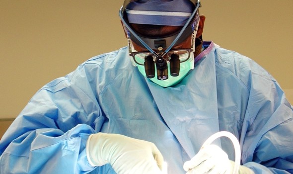
Macroscopy (gross exam)
With the macroscopic examination, it is possible to evaluate the external aspect of the animal and all its organs (feathers, ears, eyes, mouth, upper respiratory system, skeleton, body condition, etc.) with the naked eye and by touch. It is during the macroscopy that all the samples are taken for potential tests. It is always better to keep samples of each organ in formaldehyde should additional tests be necessary later on. For monetary reasons, it is sometimes impossible to perform a complete necropsy on the animal. We then have to prioritize the organs that were most likely affected according to clinical signs or organs which are visibly abnormal. A sudden death would suggest to take samples of the heart, brain, lungs and endocrine glands. In the case of a chronic disease, samples of the gastrointestinal tract and of the liver would be essential. Finally, the examination of a young bird requires to initially examine the immune system such as the bursa of Fabricius. The sampled organs are stabilized (fixed) in a formaldehyde solution. Once fixed they can be preserved for a long time.
Pictures taken at different stages of the necropsy can sometimes be very useful for a thorough investigation. In addition, X-rays can also be part of the examination in the search for fractures, tumors, heavy metals or other anomalies.
The appearance of each tissue is examined in order to determine if there is a pathological condition or not. Always following a systematical order (e.g.: preestablished form), each organ is described by its color, shape, consistency, size, smell and presence of focal lesion(s). All of these observations can be used to describe a pathological entity in the affected organ (Table # 3).
| Table 3 – Example of a macroscopic description: hepatic lipidosis (fatty liver) |
| Organ Liver |
| Colour – pale, yellow |
| Shape – rounded |
| Consistency – soft, friable |
| Size – enlarged |
| Smell – nothing to point out |
| No focal lesions |
Microscopy
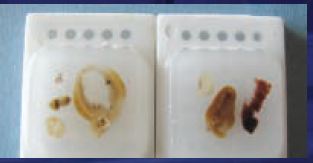
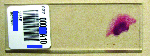
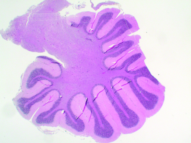
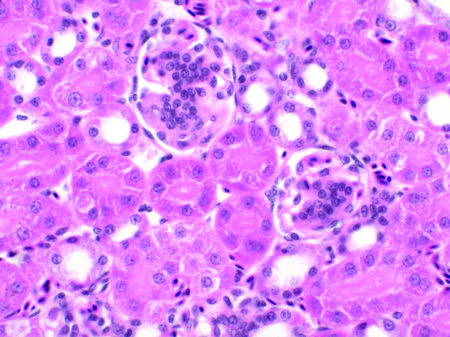
The microscopy is the study of tissues at the cellular level. To observe the cells under the microscope, many steps are necessary. In order to easily cut the tissue the water content must be removed from the cells and replaced by paraffin. Once the tissue is embedded in paraffin (image 1), it is sliced very thinly (5 microns) with an instrument called a microtome. These thin slices are then placed on a slide and stained (image 2, 3, 4). Cells are examined at different magnifications and a detailed description is made based on the histological findings – for example: inflammation, tumor, parasites, inclusion bodies etc. (Table # 4).
| Table 4 – Example of a microscopic description: splenic polyomavirus infection |
| Multifocal necrosis of the spleen tissue |
| Karyomegaly |
| Numerous basophilic intranuclear inclusions in the reticular cells of the spleen |
Ancillary Tests
The sampling of tissues, feathers or fluids for bacteriological, virological, fungal, parasitological, toxicological, special colorations, PCR tests and cytological tests are also part of the necropsy. Each test is important since it serves to complement other tests. In several cases, these tests will help to confirm the pathological diagnosis.
Conclusion
A necropsy report is based on observations done with the naked eye, under a microscope or with several types of lab tests. Of course everything that surrounds the animal is also essential. That is why all the information collected regarding the bird’s death or disease is vital. The necropsy allows us to discover much information that would not have been disclosed otherwise. This information can have a major impact on other birds. Even if a necropsy doesn’t always give a specific diagnosis, it can be the closing chapter to a probably painful event and may possibly provide a sense of satisfaction and relief. This wise choice can give us the opportunity to study a disease and then use this new knowledge to our advantage.
by: Dr. Diane Noël, D.V.M.,I.P.S.A.V
Biography – Dr. Diane Noël, D.V.M.,I.P.S.A.V
Dr. Noël graduated in veterinary medicine at the University of Montreal in 1992. She then completed an internship in small animal medicine and surgery followed by a residency in comparative pathology at the University of Miami and clinical pathology at the University of Montreal.
It is during her residency in Miami that Dr. Noël developed a growing interest in avian pathology and in the world of birds. Despite the animal diversity studied during her formal training, Dr. Noël was particularly attracted by avian hematology and the various diagnostic techniques used in avian medicine.